
In our Senology Centre in Lugano, we carry out screening and prevention examinations using mammography, ultrasound and magnetic resonance imaging.
Our specialised team guarantees professionalism and at the same time sensitivity in dealing with this special branch of medicine.
Our breast radiologists not only offer diagnostic examinations of the highest standard, but are also part of a multidisciplinary network (Swiss Cancer Screening, MIBB, EOC Centro di Senologia della Svizzera Italiana, Centro di Senologia Clinica Sant’Anna) to ensure that the patient and breast pathology are cared for from diagnosis to subsequent periodic check-ups.
Offer for examination
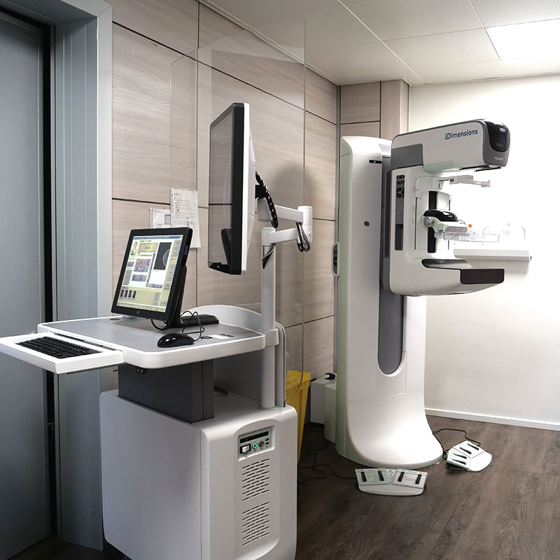
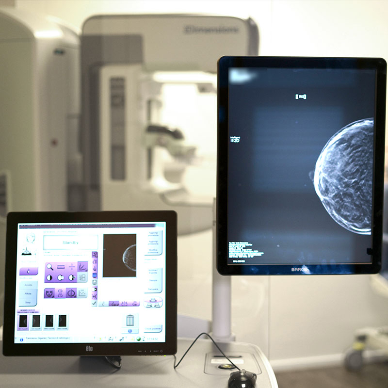
Mammography is an X-ray examination that makes it possible to examine the tissue components of the breast and detect the possible presence of non-palpable lumps and other pathologies such as microcalcifications, density asymmetries and architectural disorders at an early stage.
The cantonal mammography screening programme is a public service that offers women between 50 and 69 years of age residing in Ticino the opportunity to undergo screening mammography every two years.
Mammography as part of a high-quality organised screening programme is recommended by the Working Group of Swiss Cancer Screening Programmes swiss cancer screening, the Swiss Cancer League and the World Health Organisation as the scientifically most appropriate method for the early detection of breast cancer.
Breast tomosynthesis is a three-dimensional 3D X-ray mammography that reduces and/or eliminates the effect of overlapping tissue by taking images from different angles of a breast held still, with subsequent reconstruction of individual slices or planes at high resolution of the breast in its entirety.
Tomosynthesis is an examination that offers two basic advantages:
- False images caused by overlapping tissue layers are greatly reduced
- Reveal changes that might remain hidden with conventional mammography, even when performed digitally
The examination is combined with an ultrasound examination to better assess dense glandular tissue or to clarify doubtful findings on mammography and to differentiate possible solid/fluid lesions.
The indication for the tomosynthesis examination is given by the referring doctor and e.g. the breast radiologist:
- Patients with previous malignant breast neoplasia
- Patients with symptomatic breasts
- Patients with a high family burden of breast cancer (mother, sisters);
- Patients with a known genetic predisposition (BRCA 1 and 2 mutation);
- Women with very dense breasts with high glandular content
Breast ultrasound is a non-invasive diagnostic examination performed with an ultrasound probe. It allows the examination of the glandular tissue of the breast (i.e. the breast parenchyma) and the axilla, as well as the observation of lumps, cysts and other findings. During a biopsy, ultrasound is used to guide the needle to where the tissue is to be taken. Breast ultrasound can be performed bilaterally, when both breasts are examined, or monolaterally, when the examination is limited to the right or left breast. Breast ultrasound is an examination that, depending on the age of the patient, can detect many diseases of the breast and the lymph nodes in the armpit and is a complementary examination to mammography. It is used to examine the breasts of young women (usually up to the age of 40) whose tissue has a high glandular content, and in all those cases in
which an X-ray examination is not recommended (e.g. breast ultrasound during pregnancy). In addition, it is indicated for checking lumps that are noticed during palpation or in inflammatory diseases such as abscesses and mastitis.
In a breast biopsy, tissue is taken from the breast using ultrasound, mammography or MRI. The sample is taken from a lump or an area that has been classified as suspicious in the imaging and examined cytologically or histologically in the laboratory.
The decision on the guidance method depends on the type of lesion and its location.
Preoperative markings are methods of marking findings in the breast.
MRI of the breast is a non-invasive diagnostic examination performed with a high magnetic field (3T) machine to examine the mammary gland and lymph node stations in patients with specific indications.
The most important indications for MRI of the breast are:
- Monitoring of women at high genetic-familial risk of breast cancer, patients with a positive genetic test for a pathogenic gene mutation and women with a significant family history of breast cancer.
- Local tumour staging before surgery: this is particularly indicated in women with multiple lesions in the same breast (multifocality or multicentrality), with the contralateral breast also being examined.
- Assessment in patients undergoing neoadjuvant chemotherapy: In locally advanced breast cancer, breast magnetic resonance imaging is the most accurate technique for assessing response to treatment.
- The differential diagnosis between local recurrence and surgical scarring in operated patients when clinical examination and/or conventional imaging are equivocal or suspicious.
Book an appointment
Fill out the form and we will contact you